- Neurosurgery is a discipline that diagnoses and treats a range of injuries and disorders of the brain and the central nervous system
- For millennia the speciality was dominated by forms of craniotomies, which are procedures to remove portions of the skull to gain access to brain disorders
- In the early and mid-20th century visual, guidance and radiation technologies disrupted the treatment of some brain disorders by introducing less- and non-invasive procedures to the discipline
- At the beginning of the 21st century, a flurry of rapidly developing innovative technologies including, augmented reality, artificial intelligence (AI), robotics and genomic and cellular therapies, are accelerating the trajectory of neurosurgery towards a less- and non-invasive speciality
Brain disorders and the changing nature of neurosurgery
Populations throughout the world are growing and aging, the prevalence of age-related disabling neurological disorders is increasing, and healthcare systems are facing large and escalating demands for treatment, rehabilitation, and support services for such disorders. According to the most recent Global Burden of Disease (GBD) Study, neurological disorders are the leading cause of disability and the second leading cause of death in the world.
The total annual global burden of traumatic brain injury alone is ~US$400bn and in the US, ~16% of households are affected by brain impairment, with many individuals requiring 24-hour care. This suggests that often several family members are involved in the caregiving process, and some are juggling the responsibilities of caregiving, child rearing and employment simultaneously.
The scarcity of established modifiable risks for most of this vast and rapidly growing neurological burden suggests that innovations are required to develop efficacious prevention and treatment strategies. This Commentary describes some of these, especially those that have changed or have the potential to change neurosurgery, by making therapies less- and non-invasive, and hold out the prospect of improving patient outcomes and lowering healthcare costs.
Neurosurgery is a medical speciality concerned with diagnosing and treating a range of disorders and injuries of the brain and central nervous system (CNS) in patients of all ages. These include tumours of the brain and CNS, infections of the CNS, pituitary tumours and neuroendocrine disorders, traumatic brain injury, cerebral aneurysms and stroke, hydrocephalus and other conditions that affect the flow of cerebrospinal fluid, degenerative spine disorders, Parkinson’s disease, Alzheimer’s, epilepsy, spina bifida, and psychiatric disorders.
Treating brain conditions is complex and challenging. This is partly because the brain is one of the best protected organs of the human body. It is encased in the bones of the skull, covered by the meninges, which consist of three membranes and cushioned by cerebrospinal fluid (CSF). It is also protected by the blood-brain barrier (BBB), which is a network of blood vessels and tissue comprised of closely spaced cells, which shield the brain from toxic substances in the blood, supply brain tissue with nutrients, and filter harmful compounds from the brain back into the bloodstream. The BBB limits the ability of therapeutics to be effectively delivered to the brain and thereby complicates the treatment of CNS disorders. Further, the brain does not feel pain because there are no nociceptors (a sensory receptor for painful stimuli) located in its tissue, which often makes diagnosis of neuro disorders late when treatment becomes more challenging and costly, and survival less likely.
Such factors partly explain why neurology and neurosurgery have been slower than some other specialities to take advantage of new and evolving technologies. However, this is changing. Over the past five decades, progress in three-dimensional (3D) visualization, miniaturisation, digital technology, robotics, computer assisted manipulation, radiation therapy, early diagnosis of cancer, and precision medicine, have contributed to improvements in the diagnosis, prognosis, and prevention of some neurological conditions and started to transform neurosurgery towards less- and non-invasive procedures that efficaciously execute complex challenges, eliminate mechanistic errors, reduce operating times, and improve patient outcomes.
Further, the growing significance of applying artificial intelligence (AI) and machine learning techniques to pre-, intra- and post-operative clinical data introduces the possibility of a new suite of medical services that have the potential to enhance patient outcomes and reduce costs by improving diagnosis, planning and the rehabilitation of patients. And more recently, there are growing synergies between neurosurgery and gene and cellular therapies, which promise to accelerate personalized, non-invasive treatments for a range of neuro disorders.
In this Commentary
This Commentary is divided into 9 sections. Section 1 provides a brief history of neurosurgery, which has its genesis in ancient times when a form of craniotomy (surgical removal of a portion of the skull) was practiced and note the difference between craniotomy and craniectomy. Section 2 describes how, in the mid-20th century, neurosurgery took ~4 decades to pivot when Lars Leksell, a Swedish surgeon, introduced a stereotactic guided device that permitted the accurate positioning of probes to treat small targets in the brain, which were not amenable to conventional surgery. Shortly afterwards Leksell developed ‘stereotactic radiotherapy’, which formed the basis the Gamma Knife®, a device that provides non-invasive surgeries for a range of brain disorders. Section 3 details how advances in magnification, illumination, and the development of fibreoptics contributed to less-invasive endoscopic neurosurgeries, which facilitated a range of brain disorders to be treated through a small burr hole in the skull. Previously such procedures would have required a craniotomy. This section also notes the rapid development of endovascular neurosurgery, which uses tools that pass-through blood vessels to diagnose and treat diseases and conditions of the brain rather than using open surgery. Today, neuro-endovascular surgery is the most practiced therapeutic approach for a range of vascular conditions affecting the brain and spinal cord and is positioned to grow further over the next decade. Section 4 suggests howneurosurgery has benefitted from a range of rapidly developing 21st century technologies including: augmented reality, artificial intelligence (AI), robotics and genomic and cellular therapies. All help to increase less- and non-invasive neurosurgical procedures and contribute to advancing personalized therapies that improve patient outcomes and lower costs. Section 5 provides some insights into the life of a neurosurgeon through the lens of Henry Marsh, an English neurosurgeon who, between 2014 and 2022, published three candid memoirs, which chronicle his career, describe daily challenges and frustrations of the speciality and explain how neurosurgical units have changed the way they are organized and run. Sections 6 briefly mentions the increasing prevalence of dementias. Although outside the direct realm of neurosurgery, the scale and speed of their growth are likely to have an indirect impact on it. Section 7 introduces traumatic brain injury (TBI), a condition caused by a blow to the head and suffered by millions. The section describes the gold standard management of severe TBI and flags a pressing need to develop a non-invasive modality for managing the condition. Section 8 notes the frustration of neurosurgeons with the late diagnosis of brain tumours and describes well-resourced global endeavours to detect a wide range of cancers from a single blood test in asymptomatic people. Takeaways follow in Section 9 and suggest that a significant proportion of neurological disorders, which previously were treated with craniotomies, are now treated with either less- or non-invasive procedures. With the speed at which technology and biomedical science are developing, the only direction of travel for neurosurgery is towards non-invasive procedures.
Section 1
History
Neurosurgery has a long history with its genesis in Mayan civilizations ~1500 BCE, who practiced cranial deformations that included flattening frontal skull bones. During the Egyptian era, when mummification started to be practiced ~2,500 BCE, embalmers did not use a form of craniotomy to gain access to the brain. Instead, they used hooked instruments to remove the brain through the nose: a prototype of modern transsphenoidal surgery, which is a common procedure today for removing tumours of the pituitary gland. Rather than opening the skull with a traditional craniotomy, the physician reaches the tumour through the nasal passages and the sphenoid sinus.
In ancient Peru Inca surgeons practiced an early form of craniotomy referred to as trepanation, which used a scraping technique to penetrate the skull. Such procedures were performed on adult men to treat injuries suffered during combat. A version of this procedure called a trephination was also practiced in Egyptian and Roman times and performed on individuals who had experienced head traumas. The approach entails making a hole in the skull to relieve the build-up of intracranial pressure (ICP) caused by brain oedema (swelling) and is described by Hippocrates in the Greek era. The first known neurosurgery in Greece took place ~1900 BCE in Delphi when skull trephinations were probably performed for religious reasons. Later, the technique was recommended by Galen during the Roman period for people who had suffered a traumatic brain injury (TBI) in battle. From ~500 to ~1500 AD, the rise of religion and war resulted in many craniocerebral traumas, which contributed to the early development of neurosurgery as a distinct specialty.
Similar trephination procedures were performed during the American Revolutionary War, which secured American independence from Great Britain, and culminated in the Declaration of Independence on July 4, 1776. During the war soldiers suffered TBIs after being hit on the head with the butt of a rifle. Although the treatment for severe TBI is similar today, (see Section 7) the main difference is that the surgical instruments used in the 18th century were not powered. About 132 years later, in 1909, Theodore Kocher, a Swiss physician and Nobel Laureate in Medicine was the first person to systematically describe a decompressive craniectomy procedure for severe TBI patients. A craniectomy is different to a craniotomy. The latter is a surgical procedure in which a section of the skull is removed to expose the brain and is performed to treat various neurological conditions, or when an injury or infection has occurred in the brain. A craniectomy involves a different surgical technique and is used on people suffering severe TBI to relieve brain oedema. In such a procedure the bone fragment removed may not be replaced immediately and is either replaced during a subsequent surgery or discarded in favour of a future reconstruction using an artificial bone.
Section 2
Stereotactic surgery
For millennia, a form of craniotomy dominated what we now know as neurosurgery. During the 20th century advances in medical science paved the way for the introduction of less- and non-invasive modalities to treat brain disorders (see below). A landmark event occurred at the beginning of the 20th century with the introduction of stereotactic surgery, which makes use of three-dimensional (3D) coordinates to locate and treat lesions in the brain. The method was first reported in the May 1908 edition of Brain, by two British surgeons Victor Horsley, and Robert Clarke. The device they described became known as the Horsley-Clarke apparatus, and was used to study the cerebellum in animals by enabling accurate electrolytic lesioning to be made in the brain of a monkey. It took ~40 years before the technique was introduced to humans following the publication of a seminal paper by Ernest Spiegel and Henry Wycis, in the October 1947 edition of Science. Spiegel was a Vienna trained neurologist who moved to Temple Medical School in Philadelphia, which in 2015 was renamed the Lewis Katz School of Medicine. Wycis was one of Spiegel’s students who became a neurosurgeon. By the time they published their 1947 paper, they had performed several neurosurgeries and there had been sufficient advances in neurophysiology, pneumoencephalography, radiology, and electrophysiology for them to design a device like the Horsley-Clarke apparatus, which was fixed to a patient’s head by means of a plaster cast and was accurate enough to be used in human stereotactic surgery. Spiegel’s and Wycis’s surgical innovations attracted attention from physicians internationally, but there were no commercial stereotactic frames and neurosurgeons were obliged to design and manufacture their own. A pivotal moment occurred in 1947, when Lars Leksell, a Swedish physician and Professor of Neurosurgery at the Karolinska Institute, in Stockholm, visited Wycis in Philadelphia and afterwards designed a lightweight titanium head frame to provide the basis for stereotactic surgery, which he described in a 1949 paper entitled, ‘A stereotaxic apparatus for intracerebral surgery’.
The Gamma Knife® In the early 1950s, Leksell and Börje Larsson, a biophysicist from the University of Uppsala, Sweden, were convinced that agents other than cannulas and electrodes could be used to eradicate pathologies in the brain, and combined a source of radiation with a stereotactic guiding device. This led to the development of a non-invasive device, which Leksell used to perform the first radio-neurosurgical procedure and discovered that a single dose of radiation could successfully destroy deep brain lesions. He called this technique “stereotactic radiosurgery”, which, in 1968, led to the first stereotactic Gamma Knife® that used a focused array of intersecting beams of gamma radiation to treat lesions within the brain. Its success encouraged Leksell to use the device over the ensuing decade in functional brain surgeries to treat intractable pain and movement disorders. Leksell’s radio surgical device used Cobalt-60 (a synthetic radioactive isotope) as a radiation source. The basic physics that drives stereotactic radiosurgery today is substantially the same. It focuses ~200 tiny beams of radiation on a target in the brain with submillimetre accuracy. Although each beam has little effect on the brain tissue it passes through, a strong dose of radiation is delivered to the place where the beams meet.
Over time, the Gamma Knife® has been refined and enhanced and its efficacy and safety have been well established. Today, the Gamma Knife® provides a non-invasive operative system for a range of brain disorders, including small to medium size tumours, vascular malformations, epilepsy, and nerve conditions that cause chronic pain. Before its introduction such disorders were treated by surgeries, which involved craniotomies. In 1987, the Gamma Knife® was introduced into the US and installed at the Universities of Pittsburgh and Virginia. Although it took decades to achieve regulatory approval and be widely used throughout the world, the Gamma Knife® represents a significant technological advance in neurosurgery. Unlike craniotomies the device provides painless procedures that do not require anaesthesia, treatments take just one session, and patients can return to normal activities almost immediately. The Gamma Knife® is ~90% successful in killing or shrinking brain tumours, and today, there are ~300 Gamma Knife® sites worldwide, which each year treat >60,000 patients.
Neurosurgeon Ranjeev Bhangoo, Clinical Director for neurosurgery at King’s College Hospital, London, UK likens the Gamma Knife® to, “an umbrella, that sits above the patient’s head, rather like the old-fashioned hair dryers in women’s hair salons, but much bigger and more complex”, and stresses that the procedure, “is not painful. Forget any notion of surgery: there’s no knife, there’s no operating theatre. It’s done with the patient awake: you walk in, have your treatment, and walk out.” See videos.
 What is Gamma Knife Radiosurgery? What is Gamma Knife Radiosurgery?
 Is Gamma Knife Radiosurgery painful? Is Gamma Knife Radiosurgery painful?
Section 3
Endoscopic and endovascular neurosurgery
Neuroendoscopy Neurosurgery pivoted again in the 1990s when disorders that would normally require opening the skull began to be treated less invasively through a small burr hole. Improved magnification, miniaturization, and illumination of lenses and the development of fibre optics facilitated an endoscopic surgical procedure to treat hydrocephalus, a condition in which cerebrospinal fluid (CSF) abnormally accumulates in the brain. There is currently no prevention or cure for the condition, but it can be managed with surgery. The procedure includes creating an opening in the floor of the third ventricle using an endoscope (a thin, flexible, tube-like imaging instrument with a small video camera on the end) placed within the ventricular system through a burr hole in the skull. In the late 1990s, neuro-endoscopy expanded to treat lesions outside the ventricular system and the endoscopic endonasal approach was established as a technique that allowed surgeons to go through the nose to operate on areas at the front of the brain and top of the spine.
Since the early use of the endoscopic procedures for treating intrasellar pituitary adenomas, the approach has been expanded to treat a range of skull base lesions. Today, skull base surgery is undertaken to remove both noncancerous and cancerous growths, and abnormalities on the underside of the brain or the top few vertebrae of the spinal column. Because this is such a difficult area to see and reach, skull base surgery has been advantaged by endoscopic procedures where surgeons insert instruments through natural openings in the skull - the nose or mouth - or by making a small hole just above the eyebrow. This type of surgery requires a team of specialists that may include ear, nose, and throat (ENT) surgeons, maxillofacial surgeons, neurosurgeons, and radiologists. Before endoscopic skull base surgery was developed, the only way to remove growths in this area of the body was by making an opening in the skull. In some cases, today, this type of surgery may be still needed.
Recent advances in endoscope design have produced equipment that is smaller and more efficient, with improved resolution and brighter illumination, than earlier models. Such developments, combined with surgeon enthusiasm, have contributed to the expansion of neuro-endoscopy to treat a range of neuro disorders including intracranial cysts, intraventricular tumours, skull base tumours, craniosynostosis (a birth defect in which the bones in a baby's skull join too early), degenerative spine disease, hydrocephalus and a rare benign tumour called hypothalamic hamartoma.
Neuro-endoscopic surgery causes minimal damage to normal structures, carries a lower rate of complications, shortens hospital stays, minimizes cosmetic concerns associated with many neurosurgical conditions and improves patient outcomes. It is positioned to take advantage of further miniaturization of cameras and optical technology, innovations in surgical instrumentation design, and further innovation in navigation and robotics systems.
Endovascular neurosurgery Another innovation that has developed over the past five decades is endovascular surgery. The term ‘endovascular’ means ‘inside a blood vessel’. Endovascular neurosurgery uses tools that pass-through blood vessels to diagnose and treat diseases and conditions of the brain rather than using open surgery. The genesis of endovascular neurosurgery is credited to Professor Alfred Luessenhop, an American physician at Georgetown University Hospital in Washington DC, who, in 1964, carried out the first embolization of a cranial arteriovenous malformation and the first intracranial arterial catheterization to occlude an aneurysm. Over the past 60 years, endovascular neurosurgery has developed and has become a subspeciality. Today, >50% of cerebral aneurysms are treated through this minimally invasive approach.
Neuro-endovascular surgery has become the most practiced therapeutic approach for the majority of vascular conditions affecting the brain and spinal cord. It is used more frequently than open surgery for the management of complex vascular conditions, with high rates of safety and efficacy. The expansion of endovascular techniques into the treatment of stroke, the third highest cause of death in the US, has provided meaningful benefits to large numbers of patients worldwide. Further, with populations throughout the world aging neuro-endovascular techniques are poised to become one of the most necessary and important treatment modalities within neurosurgery.
With age our brains shrink, which causes a space to develop between the surface of our brain and its outermost covering. This increases the possibility that a knock to the head of a person >60 will result in a brain blood vessel rupturing and bleeding: a subdural hematoma. Research suggests that, “significant numbers occur after no significant antecedent trauma”, and could be the result of “an inflammatory process occurring at the level of the dural border cell”. A chronic version of this disorder can manifest itself within weeks of the first bleeding in which blood accumulates. With aging populations, chronic subdural hematoma (cSDH), is a condition predicted to become one of the most common neurosurgical conditions in the near-term future and expected to be treated with neuro-endovascular techniques.
Further, minimally invasive neuro-endovascular procedures are now commonly used to repair cerebral aneurysms, which are weak or thin spots on arteries in the brain that balloon and fill with blood. A bulging aneurysm can put pressure on brain tissue, and may also burst or rupture, spilling blood into the surrounding tissue (brain haemorrhage). Today most brain aneurysms are treated minimally invasively with neuro-endovascular techniques, which means an incision in the skull is not required. Instead, the surgeon guides a catheter or thin metal wires through a large blood vessel in the patient’s groin to reach the brain, using contrast dye to identify the problematic blood vessel. The aneurysm is then sealed off from the main artery, which prevents it growing and rupturing. In the US ~6.5m people are living with an unruptured brain aneurysm. The annual rate of rupture is ~10 per 100,000: ~30,000 Americans suffer a brain aneurysm rupture each year. Ruptured cerebral aneurysms are fatal in ~50% of cases and those who survive, ~66% suffer some permanent neurological deficit. Each year, there are ~0.5m deaths worldwide caused by brain aneurysms and ~50% are <50years.
Section 4
Evolving technologies affecting neurosurgery
At the beginning of the 21st century scientific and technological advances are again changing the face of neurosurgery. This section briefly describes four such changes.
Neurosurgery and augmented reality Neurosurgery relies on visualization and navigational technologies and makes liberal use of computed tomography (CT) and magnetic resonance imaging (MRI) scans during preoperative planning and intraoperative surgical navigation. More recently, augmented reality (AR) applications have been used to complement more conventional visualization and navigational technologies to enhance neurosurgery. AR can bring digital information into the real environment and is beginning to play an increasing role to help neurosurgeons train, as well as plan and perform complex surgical procedures. In June 2020, surgeons atJohns Hopkins University successfully carried out a spinal fusion surgery for the first time in the US using xvision™, an FDA approved AR device for spine surgery developed by Augmedics Inc., a Chicago based company, which went public in 2020 through a reverse merger with Malo Holdings. Xvision™ allows surgeons to “see” the patient's anatomy through skin and tissue as if they have X-ray vision, to accurately navigate instruments and implants during surgical spine procedures. Each year, there are ~1.62m instrumented spinal procedures performed in the US, the majority of which are undertaken using a freehand technique, which can lead to suboptimal results.
Neurosurgery and artificial intelligence Such heavy use of advanced imaging and guidance technologies creates a vast amount of clinical data during a patient’s neurosurgical journey. It is not altogether clear how effectively pre-, intra-, and post-operative clinical patient data are collected and analyzed to enhance surgical procedures and patient outcomes. An article in the August 2021 edition of the journal Neuroscienceentitled, ‘Neurosurgery and Artificial Intelligence’, suggests that the collection and analysis of such data are beginning to happen. Over the past decade, AI techniques applied to data collected during patients’ neurosurgical journeys have enhanced diagnoses and prognostic outcomes and contributed to post operative care and the rehabilitation of patients. Being able to predict prognosis, identify potential postoperative complications, and track rehabilitation are enhanced with AI applications. The future suggests that the symbiotic relationship between AI and neurosurgery, which today is in its infancy, is positioned to grow. This will not only help AI to develop better and more robust algorithms but will provide opportunities for MedTechs to gain access to new revenue streams by providing enhanced patient services.
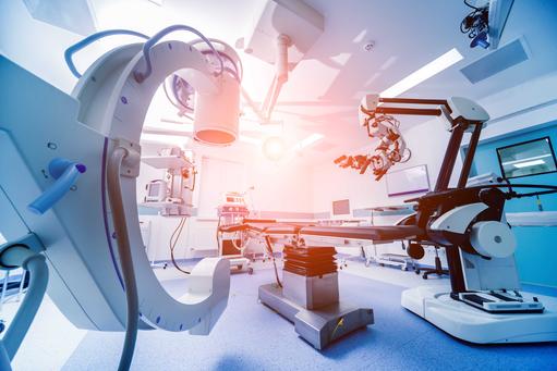
Robotics Linked to medical imaging and navigation technologies is the increasing use of surgical robotics. However, neurosurgery has been slower than other specialties to incorporate robotics into routine practice owing to the anatomical complexity of the brain and the spatial limitations inherent in neurosurgical procedures. Notwithstanding, the first documented use of a robot-assisted surgical procedure was in neurosurgery. In 1985 Yik San Kwoh and colleagues, at the Memorial Medical Center in Long Beach, California, used an Unimation Programmable Universal Machine for Assembly (PUMA) 200 (which was originally designed for General Motors’ factories) to perform a CT-guided stereotactic biopsy of a brain lesion. Although discontinued, the PUMA 200 is considered the predecessor of current surgical robots. There are now several robotic systems that have gained regulatory approval for cranial surgery. These include Zimmer Biomet’s ROSA ONE Brain, which obtained FDA approval in 2012 for intracranial applications, and Renishaw’s Neuromate robotic system, which was granted approval by the FDA in 2014. The former has been used extensively in the treatment for epilepsy, and the latter provides surgeons with five degrees of freedom for use in stereotactic applications. Robotics is a fast-moving discipline, which together with AI and machine learning, is positioned to impact neurosurgery in the near to medium term.
Neuro-pharmaceuticals and Trojan horses There are growing synergies between neurosurgery, gene, and cellular therapies. However, the BBB, which plays a significant role in controlling the influx and efflux of biological substances essential for the brain to operated effectively, makes it extremely difficult to effectively deliver drugs to the brain. Over the past three decades, many biologics (medications developed from blood, proteins, viruses, or living organisms) have entered brain and CNS clinical studies. However, they have not gained FDA approval mainly because they did not have effective mechanisms to deliver neuro-pharmaceuticals across the BBB. Instead, the clinical trials were predicated upon a variety of BBB avoidance strategies. Cerebrospinal fluid (CSF) injections are the most widely practiced approach that delivers drugs to the brain by attempting to bypass the BBB. However, this results in limited drug penetration to the brain because of the rapid export of CSF from the brain back into the bloodstream. Future drug or gene-based neuro-pharmaceuticals will need to be accompanied by advances in BBB delivery vehicles.
Currently, there are numerous scientific endeavours to devise innovative and effective ways to deliver gene therapies across the BBB to the brain. Success in this regard will mean that genomic and cellular therapies will increasingly have the potential to work synergistically with neurology and neurosurgery to provide non-invasive, personalized care for a range of brain disorders including Alzheimer’s, Parkinson’s, spinal muscular atrophy, spinocerebellar ataxia, epilepsy, Huntington’s disease, stroke, and spinal cord injury. Endeavours are underway to re-engineer biologic drugs as brain-penetrating neuro-pharmaceuticals using BBB molecular Trojan horse technologies. This approach employs genetically engineered molecular Trojan horses (proteins), which carry genes across the BBB to have a therapeutic impact on brain disorders. The future development of neuro-pharmaceuticals linked to effective means to deliver these across the BBB are together positioned to reduce the need for interventional neuro therapies, but this may take some time.
Section 5
A perspective: life as a neurosurgeon
Three memoirs by Henry Marsh, an English neurosurgeon who treated a range of brain disorders over a 40-year career at a leading neurosurgical unit in London, provide insights into the human dramas that occur in a busy modern hospital. Marsh studied Politics, Philosophy and Economics (PPE) at Oxford University before starting medical school at the Royal Free Hospital in London. In 1984, he became a Fellow of the UK’s Royal College of Surgeons and in 1987, was appointed a consultant neurosurgeon at the Atkinson Morley Regional Neurosciences Centre at St George’s Hospital in London, where he spent his entire career.
Marsh’s first book is an unflinching memoir entitled, Do No Harm: Stories of Life, Death and Neurosurgery, which was published in 2014, and describes, with compassion and candour, challenging professional experiences filled with risk and imminent death. The book opens with the sentence, “I often have to cut into the brain and it's something I hate doing.” His first operation as a neurosurgeon was to treat a cerebral aneurysm. Forty years ago, this would have required opening the skull to access the brain. The procedure had a profound impact on Marsh, who commented, “What could be finer than to be a neurosurgeon. The operation involved the brain, the mysterious substrate of all thought and feeling, of all that was important in human life: a mystery, it seemed to me, as great as the stars at night and the universe around us.”
Marsh describes the difficult decisions, which neurosurgeons and patients regularly must make that change lives forever. He recalls moments of celebration and gratification when complex operations go well, and candidly recounts some of the more undesirable outcomes and slips of the hand that result in devastating outcomes. Marsh liked working with American neurosurgeons and came to “love their optimism, their faith that any problem can be solved if enough hard work and money is thrown at it, and the way in which success is admired and respected and not a cause for jealously”. He found the attitudes of American surgeons, “a refreshing contrast to the weary and knowing scepticism of the English”. However, after visiting hospitals in the US he expressed some scepticism about “the extremes to which treatments can sometimes be pushed” and wondered whether American physicians and patients “have yet to understand the famous American dictum that ‘death is optional’, was meant as a joke”. Tellingly, Marsh notes that “sometimes doctors admit their mistakes and ‘complications’ to each other, but are reluctant to do so in public, especially in countries that have commercial, competitive healthcare systems.”
Marsh’s second memoir, Admissions: A Life in Brain Surgery, was published in 2017 two years after he retiredfrom his full-time job in England to work pro bono in Ukraine and Nepal. A documentary of his work in Ukraine, The English Surgeon, won an Emmy award. Marsh uses ‘Admissions’ to take an inventory of his life, which makes the book an even more introspective memoir than his first. He compares the challenges of working in troubled, impoverished countries like Nepal with his experience as a neurosurgeon in wealthy nations like the UK and US. The excesses of American medicine intrigued Marsh and he comments, “only in America have I seen so much treatment devoted to so many people with such little chance of making a useful recovery.” But he also expresses disillusionment with the administrative red tape in the English National Health System (NHS), which he maintains has eroded the authority and status of surgeons. In his final years working as a surgeon in St George’s Hospital in London he bemoans, “The feeling that there was something special about being a doctor had disappeared.” Marsh’s true love was patients and neurosurgery and at the end of his career, he was spending less time with patients and more time in meetings justifying his judgements and familiarizing himself with the latest UK government’s targets and edicts, which led him to say, “doctors need regulating, but they need to be trusted as well. It is a delicate balance, and it is clear to me that in England the government has got it terribly wrong”.
Marsh suggests that patients’ fear encourages surgeons to exaggerate their competence and knowledge to “shield our patients from the frightening reality they often face”. Over time, Marsh suggests, surgeons tend to believe the exaggerated versions of themselves. But the best un-learn their self-deception and come to accept their shortcomings and learn from their mistakes. “We always learn more from failure . . . . . . Success teaches us nothing,” Marsh writes.
Marsh’s third memoir,And Finally: Matters of Life and Death, was published in August 2022 and is very British, full of self-deprecation and dominated by the news that he is diagnosed with incurable prostate cancer. Marsh describes the sudden reversal of roles, from omniscient and omnipotent neurosurgeon to humble patient and provides descriptions of the ebbs and flows of his therapeutic journey, which gives valuable insights into how medicine in England works.
All three books bear witness to the fact that neurosurgery is a stressful and demanding profession, which requires extensive training, stamina, a high degree of manual dexterity, excellent hand-eye coordination, exquisite precision, extraordinary attention to detail, an ability to rapidly gather and process complex information to resolve challenging problems, compassion and empathy for patients, communication skills and teamwork. Unlike other surgical disciplines, a relatively small mistake can lead to “appalling disability”, coma, and death. According to research published in the October 2014 edition of Surgical Neurology International, ~25% of neurosurgical errors can be prevented or reduced with the increased use of evolving technologies, some of which are described in this Commentary.
Changes in the organization of neurosurgical units During Marsh’s 40-year career there were changes in the way neurosurgical units were organized and run; particularly the development of subspecialities among physicians and the use of multidisciplinary team approaches to clinical challenges. Much of Marsh’s career reflected a time when neurosurgeons worked in relative isolation and treated a wide range of neurosurgical conditions that presented in their clinics. Today, most neurosurgeons have a primary interest in a subspeciality such as epilepsy, neurovascular surgery, spinal surgery, the excision of tumours etc., and a secondary interest, which they share with colleagues. This tends to facilitate cross referral of patients among a team of physicians and improves patient care and the training of health professionals. In the operating room (OR) neurosurgeons work with other physicians, anaesthetists, trainee doctors, theatre nurses, and medical students. Outside the OR they collaborate with radiologists who use a range of diagnostic tools, including CT, MRI scans, and cerebral angiographies, which are used to detect abnormalities in blood vessels such as aneurysms, blockages, and bleeding. These neuroimaging technologies and neurosurgery have become inseparable. Neurosurgeons also work with neurologists, oncologists, ophthalmologists, and paediatricians. In 2017, Bob Carter, head of neurosurgery at the Massachusetts General Hospital, in the US, appreciated the interconnections between several clinical disciplines that care for people with neurological disorders and merged his neurosurgery department with the departments of neurology, psychiatry, and neuroradiology. While sub specialisms and teamwork have made an impact on the organization of neurosurgical units, new and emerging technologies have expanded the repertoire of neurosurgeons.
Awake brain procedures Marsh specialised in operating on the brain while the patient is awake. This aspect of his work was the subject of a BBC documentary, Your Life in Their Hands. Awake brain procedures are usually performed when a lesion is located near the frontal lobes responsible for motor skills and speech. In the video below, Ranjeev Bhangoo describes the procedure, “It’s a technique where the patient is awake during the brain surgery. The patient is neither in pain nor suffering. When we make a cut in the skin and raise a trapdoor in the skull the patient is completely asleep. We wake them up after that point and the good news is the brain itself doesn’t feel pain. So, you can do this operation without the patient being in any distress or pain. It’s an unusual situation and the patient is prepared for it beforehand. The reason why you might want to do an awake craniotomy is because in some situations, tumours are close to critical structures of the brain that control speech or movement. While we have good maps of the brain and we have image guidance, they’re not precise enough. You want the patient to be talking to you and you want to be stimulating bits of the brain to see precisely where speech is so that you can avoid those areas and do the same with movement, you want to see the patient moving his or her arm or leg while you’re stimulating bits of their brain. So, we use an awake craniotomy when we’re operating near to what we call ‘eloquent’ areas of the brain that, if damaged, would produce a devastating deficit such as problems with speech or movement”. See video.
 When and why is an awake craniotomy performed? When and why is an awake craniotomy performed?
Section 6
The increasing burden of dementias on healthcare systems and economies
As populations age and live longer so dementia conditions increase. Alzheimer's, which effects parts of the brain that control thought, memory, and language, is the most common dementia in Western societies. It is a progressive disorder that begins with mild memory loss and leads to a loss of the ability to carry on a conversation and respond to your environment. In the three decades between 1990 and 2019, the global incidence of Alzheimer’s and other dementias increased by ~148%. In 2022, there were >6.5m Americans living with the condition: ~73% >65 and ~66% of these women, but this simply may be due to women living longer. By 2050, it is projected that ~13m Americans will suffer from dementia, which is expected to kill 1 in 3 seniors; that is more than breast and prostate cancers combined.
According to the World Health Organization (WHO), there are ~55m people with dementia globally, and >60% are living in low- and middle-income countries (LMIC). Age is the most significant risk factor: the likelihood of Alzheimer’s doubles every 5 years after you reach 65. But also, dementias appear to be increased by conditions that damage the heart and blood vessels, which include heart disease, diabetes, stroke, high blood pressure and high levels of cholesterol. As the proportion of older people in populations increase in nearly every country, people living with dementias are expected to rise to ~78m by 2030 and 139m in 2050. There is no cure for Alzheimer's, and treatments tend to fall to neurologists. Drug therapies include galantamine, rivastigmine, and donepezil, which are cholinesterase inhibitors (also known as anti-cholinesterase, are chemicals that prevent the breakdown of the neurotransmitter acetylcholine) that are prescribed for mild to moderate Alzheimer's symptoms and may help reduce or control some cognitive and behavioural symptoms. Also, there are non-drug options. Although outside the direct realm of neurosurgery, the scale and speed of the growth of Alzheimer’s and other dementias are likely to indirectly impact neurosurgery by increasing the burden on over-stretched healthcare systems. Under such circumstances, it seems reasonable to assume that there will be increased pressure on neurosurgery to become less resource intense, which means less invasive and less costly while improving patient outcomes.
Section 7
Traumatic brain injury
On Thursday 29th September 2022, Tua Tagovailoa, the Miami Dolphins’ quarterback received a head injury during a match against the Cincinnati Bengals and was stretchered off. Four days earlier he left the field after receiving another head injury while playing against the Buffalo Bills. He was then checked for a concussion and cleared and came back onto the field in the third quarter. Subsequently, the NFL Players Association exercised its right to remove the independent neurological expert who was involved in the decision to clear Tagovailoa to return to the Buffalo Bills game after being evaluated for a traumatic brain injury (TBI). This raises the significance of injuries to the brain and the challenges of accurately assessing their severity and adequately treating them. TBI is as an alteration in brain function pathology by a sudden trauma, causing damage to the brain. Each year, the condition affects ~69m individuals worldwide. Symptoms can be mild, moderate, or severe, depending on the extent of the damage: annually ~5.5m severe cases are recorded globally. The epidemiology of the disorder is challenging because, in low-resourced regions of the world, where the prevalence of TBI is believed to be high, data are poor. According to the World Health Organization (WHO), ~90% of deaths due to head injuries occur in low- and middle-income countries (LMICs), where ~85% of the global population live and where the standards of care are patchy. TBI not only causes health loss and disability for individuals and their families, but also represents a costly burden to healthcare systems and economies through lost productivity and high healthcare costs. The total annual global burden of TBI is ~US$400bn. Since the beginning of the 20th century, our knowledge and understanding of the pathophysiology of brain oedema (swelling) in head trauma patients has increased and today decompressive craniectomy is a recognised procedure for severe TBI to mitigate intracranial hypertension and its impact on clinical outcomes. One of the largest clinical studies, which sought to determine the efficacy of decompressive craniectomies for TBI patients, was the RESCUEicp trial: findings of which were published in the September 2016 edition of the New England Journal of Medicine. The study was carried out over a 10-year period, between 2004 and 2014, on 408 randomly assigned patients, 10 to 65 years of age, and concluded that, “At 6 months, decompressive craniectomy in patients with traumatic brain injury and refractory intracranial hypertension resulted in lower mortality and higher rates of vegetative state, lower severe disability, and upper severe disability than medical care”. In the US, TBI is a leading cause of death and disability. Each year, ~1.5m Americans sustain a TBI, ~50,000 die from the insult, ~230,000 are hospitalized and survive, and ~90,000 experience the onset of long-term disability. According to the US Centers for Disease Control and Prevention, ~5.3m Americans (~2% of the population) are living with disability as a result of a TBI. In 2010, the economic impact of TBI in the US was estimated to be ~US$77bn in direct and indirect costs. Each year in the UK ∼1.4m patients attend hospital following head injury and TBI is the most common cause of death for people in the UK <40 years. Gold standard monitoring of intracranial pressure There is no cure for severe TBI, and the gold standard management is to monitor intracranial pressure (ICP), caused by brain oedema (swelling). Current clinical guidelines for raised ICP levels suggest thresholds, usually between 20 and 25 millimetres of mercury (mmHg), at which treatment is recommended to either prevent or reduce further damage to the brain. The device used to monitor ICP is an intraventricular catheter system that requires drilling a burr hole in the skull to insert a catheter and placing it in a cavity (ventricle) in the brain, which is filled with cerebrospinal fluid (CSF). This is then connected to an extra-ventricular drain (EVD) that measures ICP. Such systems are accurate and reliable, but also, they are health-resource-intensive modalities, which run a risk of haemorrhage and infection. Challenges with gold standard monitoring According to research findings published in the January 2017 edition of the Journal of Neurosurgery, haemorrhage is a common complication of an EVD placement. Among the cases in which patients underwent imaging after a placement procedure, haemorrhage was found in 94 (21.6%). Another study, of 246 EVDs placed in 218 patients over a 30-month period and published in the November 2014 edition of Interdisciplinary Perspectives on Infectious Diseases, reported the cumulative incidence of EVD-related infections to be 8.3%. Further, because of the dearth of qualified neurosurgeons in under-resourced regions of the world, EVD systems are not widely available in LMIC, where the incidence rates of TBI are understood to be high and increasing. Non-invasive ICP monitoring Numerous alternatives to invasive gold standard ICP monitoring are in development, but none have established a valid place within a daily clinical setting. A review paper published in the December 2020 edition of the journal Neurotrauma, entitled “Non-Invasive Techniques for Multimodal Monitoring in Traumatic Brain Injury: Systematic Review and Meta-Analysis”, stresses the significance of monitoring ICP and brain oxygenation continuously in severe TBI patients, and suggests that the “two most prominent and widely used technologies for non-invasive monitoring in TBI are near-infrared spectroscopy [a form of photoplethysmography (PPG)] and transcranial Doppler”. Researchers conclude that, “both techniques could be considered for the future development of a single non-invasive and continuous multimodal monitoring device for TBI”. Transcranial Doppler (TCD) ultrasonography is a non-invasive, painless ultrasound technique that uses high-frequency sound waves to measure cerebral blood flow velocity that may correlate with ICP. Research suggests that in ~15% of cases the ultrasound waves are unable to penetrate the patients’ skulls, and measurement is prone to intra- and inter- observer variability and accuracy. As the TCD system for measuring ICP non-invasively is encountering challenges, so near infra-red spectroscopy is gaining significance. This is a form of PPG technology, which is an uncomplicated, inexpensive, non-invasive, and convenient optical measurement that has the potential of being used at the site of injuries to quickly assess the severity of the head trauma. In the recent case of Tagovailoa, such a non-invasive ICP measurement device could have been applied on the playing field. Over the next decade, expect PPG technology to impact neurosurgery by potentially providing more accurate triaging and further disrupting the gold standard of care for severe TBI patients.
Section 8
Brain cancer and early diagnostics
We mentioned the Gamma Knife’s® ability to treat some brain tumours and suggested that patients have benefitted significantly from its use. The first successful modern brain tumour excision was performed in 1878 by William Macewen, a pioneering Scottish surgeon, at the Glasgow Royal Infirmary. At the beginning of the 20th century, contributions by Americans started with Harvey Cushing, who is generally recognised as the father of modern neurosurgery. Working at the John’s Hopkins Hospital in Baltimore, Cushing introduced meticulous documentation of the clinical and pathological details of cerebral tumours and devised several surgical techniques for operating on the brain that became the foundation of neurosurgery as an autonomous surgical discipline. In 1912, he discovered an endocrinological syndrome caused by a malfunction of the pituitary gland, which is named after him: Cushing’s disease.
The prognosis for a brain tumour is dependent upon its type, location, size and time of diagnosis, growth and how much can be surgically removed or treated. Factors including age and general wellbeing as well as some recognised genetic factors also influence prognosis. Poor prognosis for brain cancers is perpetuated by the lack of cost-effective, accurate tests that can be used in a primary care setting to diagnose the conditions. This means that a large proportion of brain cancers are diagnosed too late for current treatments to be effective. However, in recent years there have been advances made in detecting brain cancers early and this is expected to significantly improve prognosis.
Although there are >120 different types of brain tumours, lesions and cysts, your chances of developing brain cancer is <1%. Brain tumours account for ~90% of all primary central nervous system (CNS) tumours. In 2020, >0.3m people worldwide were diagnosed with a primary brain or spinal cord tumour. According to the Annual Report of the US Central Brain Tumor Registry, >84,000 Americans were diagnosed with a primary brain tumour in 2021. The US National Cancer Institute, suggests ~0.6% of Americans will develop brain cancer in their lifetime and the 5-year survival rate for those that do is only ~33%. This year, >4,000 Americans <15, are expected to be diagnosed with a brain or CNS tumour. In the UK, each year ~16,000 people are diagnosed with a brain tumour and ~ 60,000 people are living with a brain tumour.
The causes of brain tumours are not fully understood and occur because of an abnormal growth of brain cells or cells in the brain’s supporting tissues, which can damage the brain, threaten its function and result in death. Some tumours may occur around the edge of the brain and press on certain parts of it, while others can be more diffuse and grow among healthy tissue. In the video below, neurosurgeon Christopher Chandler, who leads the Paediatric and Adolescent Neurosurgical Service at King’s College Hospital, London explains that, “A brain tumour is an uncontrolled growth of a bunch of cells where the ‘off’ switch is missing. This means that there’s nothing telling these cells to stop growing, so they grow and divide. As this uncontrolled mass, or tumour, grows it displaces brain tissue and causes pressure on the surrounding brain. If you don’t remove the tumour or stop it from growing, it will grow so large that it causes critical pressure on the surrounding structures of the brain, which eventually, if untreated, can kill the patient.” See video.
 What is a brain tumour?
The Holy Grail Neurosurgeons are frustrated by the fact that brain cancers are often diagnosed late. This is because brain tumours often present with non-specific symptoms and are therefore challenging to diagnose. In the video below, neurosurgeon Ranjeev Bhangoo explains the reasons for a brain tumour to be diagnosed late. “Firstly, the symptoms are non-specific: tiredness, headache, poor concentration - maybe not finding your keys as well as you use to – the sort of thing that can happen to any of us when we’re tired. The classic thing of having a fit or collapsing occur, but they’re unusual. Your GP is only likely to see just one or two brain tumour cases in his or her whole career. . . . Now, if you do get a scan, the chances of you having a brain tumour are incredibly rare. So, just because a neurologist has organized a scan, you mustn’t get worried because it’s very unlikely that you’ll have a brain tumour. But ultimately, through some path or other, you have a scan, usually a CT scan, which is a form of X-ray, which is quick and safe and if there is a tumour it will show. At that point, what will normally happen is that your doctor will refer you to a neurosurgeon”.
 How are brain tumors diagnosed?
Technologies positioned to reduce neurosurgeons’ frustration with late diagnosis of brain cancers are quick, easy-to-use, and inexpensive blood tests that can diagnose cancer early. Such tests fall into four general categories: (i) complete blood count used to evaluate your overall health and detect a wide range of disorders, (ii) biomarkers, which are molecules found in your blood and other body fluids that can indicate specific cancers, (iii) blood protein testing that measures the amount of protein in your blood to diagnose cancer, and (iv) circulating tumour cell tests, which look for tumour cells that are shed from a tumour and are now circulating through your bloodstream.
Detecting brain cancers early Two recent examples of simple diagnostic blood tests are reported in the August 2022 edition of Clinical Cancer Research and the October 2019 edition of Nature Communications. In the former paper, scientists at Massachusetts General Hospital (MGH) report findings of a study, which detected the presence of brain cancers early by identifying pieces of tumour cells’ genetic material - mRNA - that circulate in your blood. The test, which has a sensitivity of 72.8% and a specificity of 97.7% can characterize brain tumours and monitor their status after treatment. According to Leonora Balaj, a co-senior author, and assistant professor of Neurosurgery at Harvard Medical School, “There is a real need to make brain tumor diagnosis less invasive than the current technique of tissue biopsy. This research demonstrates that it is now feasible to diagnose a brain tumor via a blood test for one of the most common mutations detected in brain tumors”. Findings of the latter paper suggest that certain brain cancers may be detected early from a simple blood test using PPG technology, which has been used in hospital settings since the 1980s to monitor heart rate and relative blood volume. Today, the technology is used in a wide range of commercially available medical devices, as well as smartwatches (the Apple version is an FDA approved medical device) and fitness trackers, for measuring oxygen saturation, blood pressure and cardiac output, assessing autonomic function and detecting peripheral vascular disease. Previously we described how PPG technology is positioned to provide a non-invasive means to monitor ICP in TBI patients.
The 2019 Nature paper describes how PPG easily, cheaply, and accurately identified asymptomatic people with suspected brain cancer. In the first instance, the technology was used on a retrospective cohort of 724 people, which included those with primary and secondary cancers as well as control participants without cancer. PPG was employed to identify biomarkers from patients’ blood samples and a machine learning algorithm was trained to identify specific biomarkers with cancer present. The algorithm was then used on a sample of 104 random participants and brain cancer was detected in 12. The PPG test revealed a sensitivity of 83.3% and a specificity of 87%. According to Matthew Baker, from the University of Strathclyde, Scotland, the paper’s lead author, “This is the first publication of data from our clinical feasibility study, and it’s the first demonstration that our blood test works in the clinic.”
A global endeavour These two studies are part of a well-resourced global endeavour to develop an affordable, simple, point-of-care, blood test, which detects cancer before any symptoms occur. Today, biomedical advances move at a much faster pace than medical technology did in the 1950s and 60s when Lars Leksell was developing minimally invasive stereotactic radiosurgery procedures to accurately locate and remove brain tumours. For example, in ~7 years since its foundation in 2015, GRAIL, a US biomedical start-up backed by Jeff Bezos and Bill Gates, has become a global leader in a ground-breaking multi-cancer, early detection, blood test, Galleri®, which has the potential to detect >50 types of cancers before they are symptomatic. This is achieved by looking for abnormal DNA shed from cancer cells in the blood, called cell-free DNA (cfDNA). The Galleri® test uses genetic sequencing technology and artificial intelligence (AI) to scan for patterns of chemical changes in the cfDNA that come from cancer cells but are not found in healthy cells. If validated, the GRAIL test will provide a simple, cheap, non-invasive means to identify a range of cancers in asymptomatic people when they are more likely to respond positively to therapy.
Large UK clinical study In May 2019, the GRAIL Galleri ® blood test was granted US FDA Breakthrough Device designation. The test is only available commercially in the US but is rapidly gaining provenance in other regions of the world. For example, in September 2021, NHS England launched a massive clinical study for Galleri® and set up ~150 mobile clinics in convenient locations across the country to recruit ~140,000 participants. In July 2022, participants were invited to attend two further appointments spaced ~12 months apart. Findings from the study are expected to confirm the accuracy of the test in asymptomatic participants and lead to its regulatory approval. Although Galleri® is the first of its kind to be trialled on such a scale in the UK, it is not the only player and cfDNA is not the only technology.
Guardant Health Another US biotech developing capabilities to detect a range of cancers early from a simple blood test is Guardant Health. Founded in 2011, the company is now ~US$6bn Nasdaq traded global enterprise with annual revenues ~US$110m. In April 2022, Guardant presented new data at the American Association for Cancer Research Annual Meeting. Findings suggested that the company’s investigational next-generation Guardant SHIELD™ assay has the capacity to analyse ~20,000 epigenomic biomarkers that help to detect a broad range of solid tumours using a single blood test. Guardant’s co-CEO, Amir Ali Talasaz said: “These positive results show that the next-generation Guardant SHIELD multi-cancer assay provides sensitive detection of early-stage cancers with the ability to identify the tumor tissue of origin with high accuracy”.
Section 9
Takeaways
For millennia neurosurgery, which has its roots in ancient civilizations, was dominated with forms of craniotomies, which opened the skull to access cerebral disorders. In the 20th century the speciality pivoted and introduced less- and non-invasive procedures to deal with a range of brain and CNS conditions. However, the introduction of these were slowed by the fact that the brain is such a well-protected organ and they took nearly half a century to gain regulatory approval and enter the clinic. At the beginning of the 21st century biomedical research is advancing at such a pace and it positioned to significantly transform neurosurgery towards a less- and non-invasive modality. Further, in the next two decades expect gene and cell therapies to substantially increase their influence as treatments for neurodisoders. Over the past three decades novel neuro-pharmaceuticals have been constantly in clinical trials but failed to receive regulatory approval because they did not have an efficatious mechanism to deliver the therapeutics across the BBB. Today, there are a myriad of novel vehicles under development, which are expected to effectively smuggle 21st century pharmaceuticals across the BBB. These are being advanced in parallel to the drugs, and together are positioned to significantly disrupt traditional neurosurgical procedures over the next two decades.
|